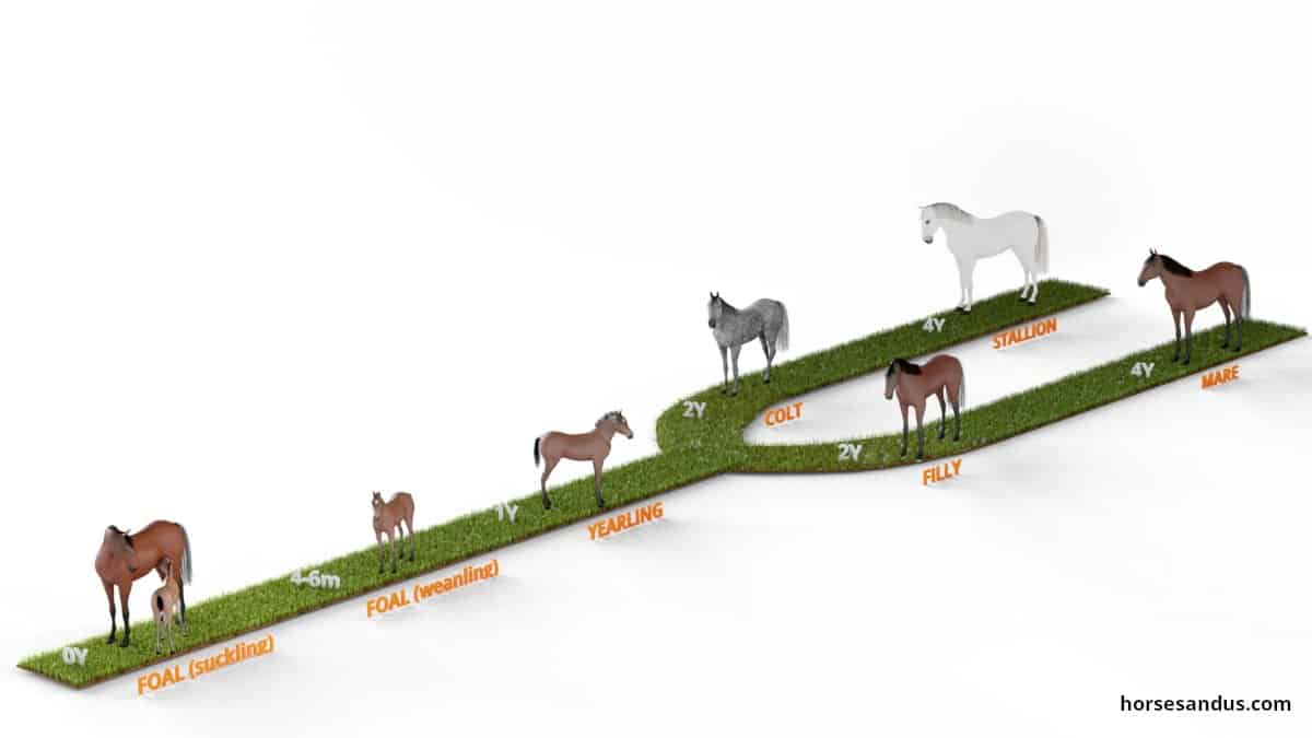
Special attention should be given to a foal’s growth rate and skeletal development.
Developmental Orthopedic Disease (DOD) in horses is a wide range of bone and joint abnormalities associated with the skeletal growth process. These growth disorders generally include osteochondrosis, physitis, angular limb deformities, flexural limb deformities, and Wobbler Syndrome.

These diseases appear when there is a disruption in the equine equine skeletal development. More specifically, in the physiological process that forms cartilage and converts it into bone.
This process occurs in the cartilage of the physis (growth plate) and the articular cartilage of the joint.
Developmental Orthopedic Disease (DOD) in horses is multifactoral, having several possible causes:
- overly rapid growth
- inadequate diet
- excessive exercise
- hormonal diseases
- genetic predisposition.
Although the incidence of these problems is high (10% to 50%), many of these conditions can be treated with changes to diet and stud management.
Any horse can have Developmental Orthopedic Disease, although ponies and feral horses are much less affected.
Let’s take a look at some of these conditions.
Equine Osteochondrosis
Equine Osteochondrosis is one of the most common developmental orthopedic disease in horses, and its incidence appears to be increasing. It occurs worldwide in many breeds costing the horse industry enormous amounts of money.
This condition involves damage to the growth cartilage due to failure of blood supply. It mainly affects the articular cartilage (immature joint cartilage covering the ends of the bone) and underlying bone.
However, it can also affect cartilage within the growth plates. In this case, bone shape and length can be disturbed.
Symptoms usually start their appearance in yearlings who have just initiated their training. Typically they will present a swollen joint which can be accompanied by a mild lameness.
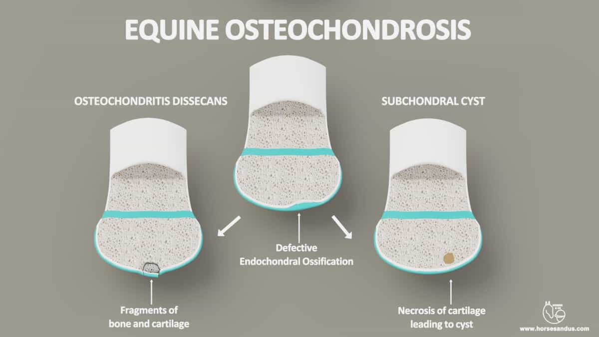
Characteristic Manifestations of Equine Osteochondrosis
Equine Osteochondrosis has two characteristic manifestations:
1 – Osteochondritis dissecans. A defect in endochondral ossification causes the thickening of joint cartilage with subsequent cracking, breaking into loose cartilage pieces called joint mice. The detached pieces can lead to joint inflammation and lameness.
2– Subchondral bone cysts. A defect in endochondral ossification causing the formation of cartilage pockets retained deep beneath the joint´s surface and its subsequent necrosis leading to the formation of cysts.
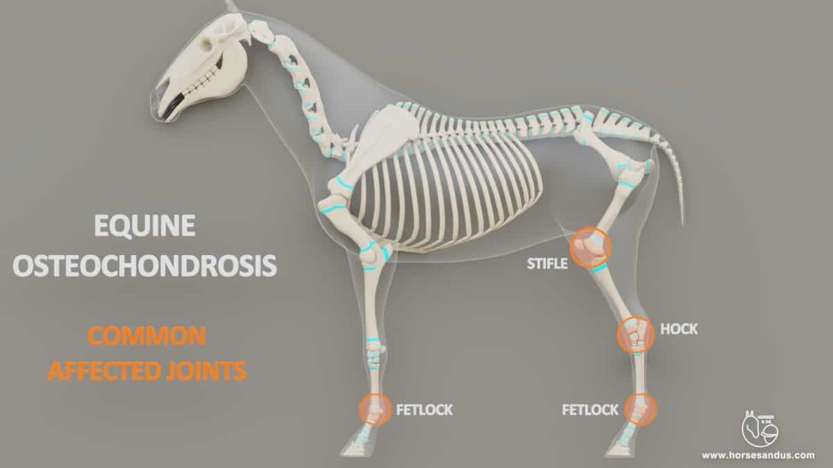
Which Joints Can Be Affected by Equine Osteochondrosis?
Equine Osteochondrosis can appear in any joint of the horse’s skeleton. It most frequently occurs in the hock, stifle, fetlock. However, it can also appear in the neck, shoulder, elbow, and hip.
Causes of Osteochondrosis in Foals
Osteochondrosis can have several causes:
- Nutrition is the most important cause. Either excess or deficiency of nutrients.
- Fast growth rates.
- Excessive exercise may cause traumatic damage to articular cartilage.
- Genetic predisposition
Treatment of Osteochondrosis in Foals
The treatment will depend on the severity and the location of the problem.
Mild cases can recover without treatment or with a restricted diet to slow down the growth rate and reduced exercise to avoid more stress to the joint. Intra-articular medication to reduce inflammation may also be beneficial.
More severe cases may require surgery to remove cartilage and bone fragments, and trimming the ragged articular cartilage.
(sources of equine Osteohondrosis used for this article: sciencedirect, thehorse, vcahospitals)
Equine Physitis
Physitis means an inflammation of the growth plate (physis = growth plate, itis = inflammation).
This condition can also be called Physeal Dysplasia which is probably a more appropriate term since in the initial stage of the disease there is no active inflammation.
Physitis is sometimes referred to as ‘“growing pains”
Physitis is a developmental Orthopedic disease in horses caused by a disturbance of the bone growth process. It is characterized by an enlargement of the growth plate which creates a flaring at the end of the bone, giving it the shape of an hourglass.
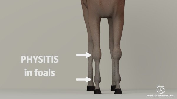
Young horses with physitis can be very lame or just show some stiffness when they are moving. Sometimes the signs are almost unnoticeable such as the foal not running as much as usual. In more severe cases the area around the growth plate may be swollen, warm and painful.
This condition only affects young growing horses while the growth plates on the long bones are still not closed.
Which Bones Can Be Affected by Equine Physitis?
The bones commonly affected are the radius, tibia, and cannon bones. More specifically, the distal (lower) growth plates of these bones are affected.
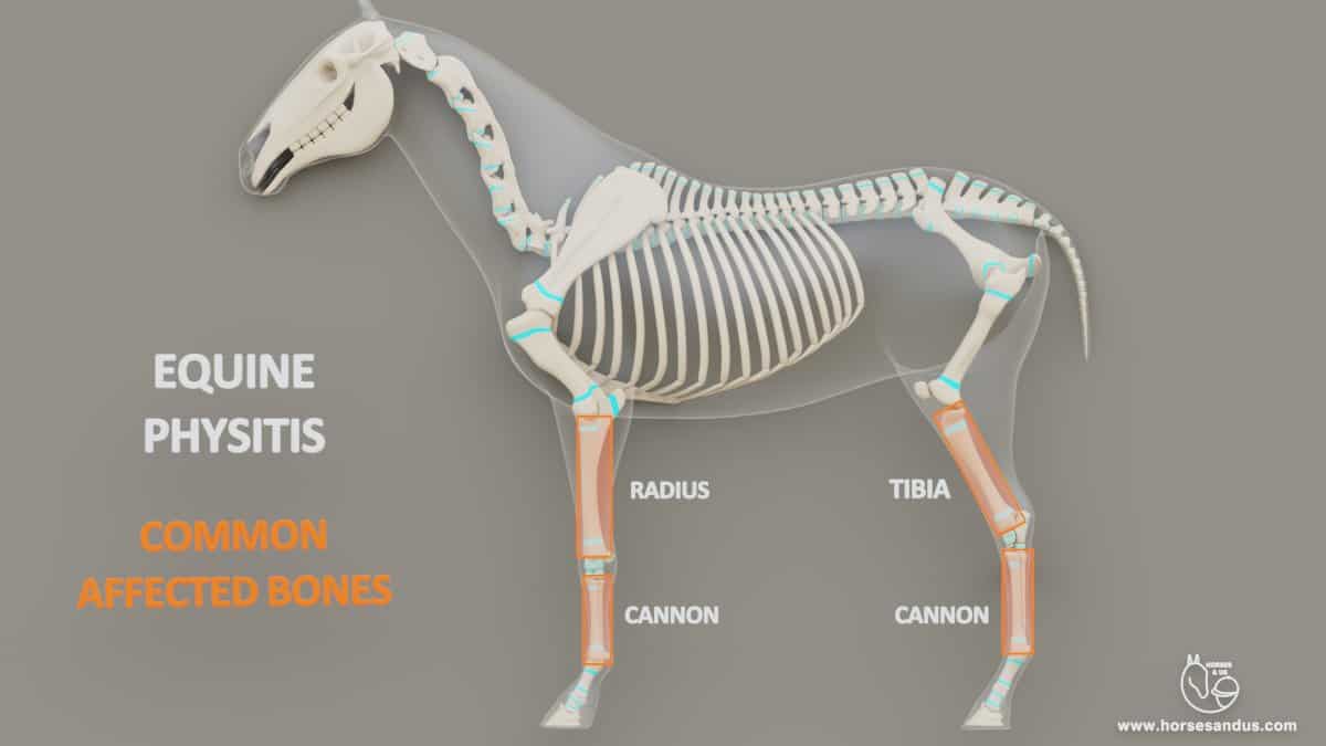
Physitis usually only occurs in one or two limbs but occasionally all four limbs can be affected.
Causes of Physitis in Foals
Physistis can have several causes:
- Nutrition is the most important cause, either excess or deficiency of nutrients.
- Trauma or excessive weight-bearing on a certain growth plate. This can be caused by excessive exercise or limb deformities that create uneven weight-bearing.
- Genetic predisposition
- Infectious cause. A growth plate can also become inflamed due to a bacterial infection.
Treatment of Physitis in Foals
The treatment of physitis consists mainly in two components:
- Rest will limit exercise and, therefore, the compression of the physis when the foal is moving.
- Restriction of diet, so the foal reduces body weight and delays the excessive growth speed limiting it to a normal rate.
Additionally the veterinary may also recommend some medication for the pain and inflammation.
(sources of equine physitis used for this article: aaep, thehorse, ker, foranequine)
Equine Angular Limb Deformities (ALD)
Angular limb deformities are classified as Developmental Orthopedic Disease. These deformities are common in foals, and a large proportion will naturally correct within a week or two of life. The more severe cases can usually be corrected with appropriate and timely treatment.
A foal has an angular limb deformity when his legs are bent either outwards (valgus) or inwards (varus).
The most common joint affected is the carpus (knee) of the foal. However, the fetlock (ankle) and tarsus (hock) can also be affected. Usually, more than one leg is affected.
The Various Equine Angular Limb Deformities (ALD)
The Angular limb deformity is named according to the joint and the direction in which the leg is bending.

Carpal Valgus commonly named “knock-kneed.” The limb deviates outwards below the knee. This is the most common form of angular deformity.

Carpal Varus commonly named “bow legs.” The limb deviates inwards below the knee.

Fetlock Valgus commonly named “toe-out.” The limb deviates outwards below the fetlock.

Fetlock Varus commonly named “toe-in” or “pigeon-toed.” The limb deviates inwards below the fetlock.

Tarsal Valgus commonly named “cow hocked.” The limb deviates outwards below the hock
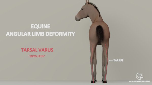
Tarsal Varus commonly named “bow legs.” The limb deviates inwards below the hock.

Windswept is a deformity where one limb is bent outward (valgus), and the opposite limb is bent inward (varus)
How Equine Angular Limb Deformities Are Developed
Any disturbance to the endochondral ossification process (bone growth) can lead to developmental orthopedic disease. When this disturbance occurs in the physis (growth plate), or the small bones of the carpus (knee), or tarsus (hock), it can lead to Angular Limb Deformities.

1- Disturbance in the Physis (growth plate)
- All sides of the physis should develop at the same rate. When there is an unequal growth across the physis, the bone will not grow straight, and an angular limb deformity will occur.
2- Disturbance in the small bones of the carpus(knee) or tarsus(hock)
- When the small bones do not complete the ossification process, they are soft and can easily crush under pressure. As a result, the neighboring long bones will fall into an angular position which results in an angular limb deformity.
Causes of Angular Limb Deformities in Foals
The cause of these deformities is multifactorial. The two main categories are congenital (present at birth) and developmental (acquired after birth).
Congenital– Maternal diseases or premature birth can result in incomplete ossification of the small bones in the knees and hocks.
Developmental – after the foal is born, he can develop these deformities due to inadequate nutrition, excessive exercise, or lameness in the opposite limb.
Treatment of Angular Limb Deformities
The treatment depends on the degree of the deformity.
Mild angular deformities generally do not require any treatment, or they can be resolved with corrective trimming and shoeing with hoof extensions.
For more severe cases surgical treatment is required.
(sources of equine angular limb deformities used for this article: ker, rossdales, dvm360, researchgate).
Equine Flexural Limb Deformities (FLD)
Flexural limb deformity is a limb deviation where one or more joints have a hyperflexion (bent backward) or a hyperextension (bent forward) when seen from the side.
Flexural limb deformities are a Developmental Orthopedic Disease in horses. These deformities affect the tendons (unlike angular limb deformities, which affect the bones).
Hyperflexion is usually described as “contracted tendons” although this is incorrect because the tendons are not actually contracted. They are too short in relation to the skeletal structure.
Hyperextension is often called laxity or “lax tendons”.
The most commonly affected joints are the distal interphalangeal joint (coffin), metacarpophalangeal joint (fetlock), and carpus joint (knee).
These deformities are much more common in the forelimbs than the hindlimbs.
The Various Equine Flexural Limb Deformities (FLD)
The different forms of flexural deformities are as follows:
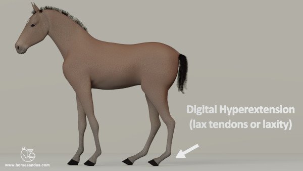
Digital Hyperextension is nearly always congenital, and many foals are born with some degree of digital hyperextension. It’s caused by flaccid flexor muscles. It is also referred to as lax tendons or laxity.
This deformity can range from slightly dropped fetlocks to fetlocks actually touching the ground. In more severe cases the toes lift off the ground.
In mild cases, the deformity corrects itself as the flexor muscles strengthen. However more severe cases will require treatment.
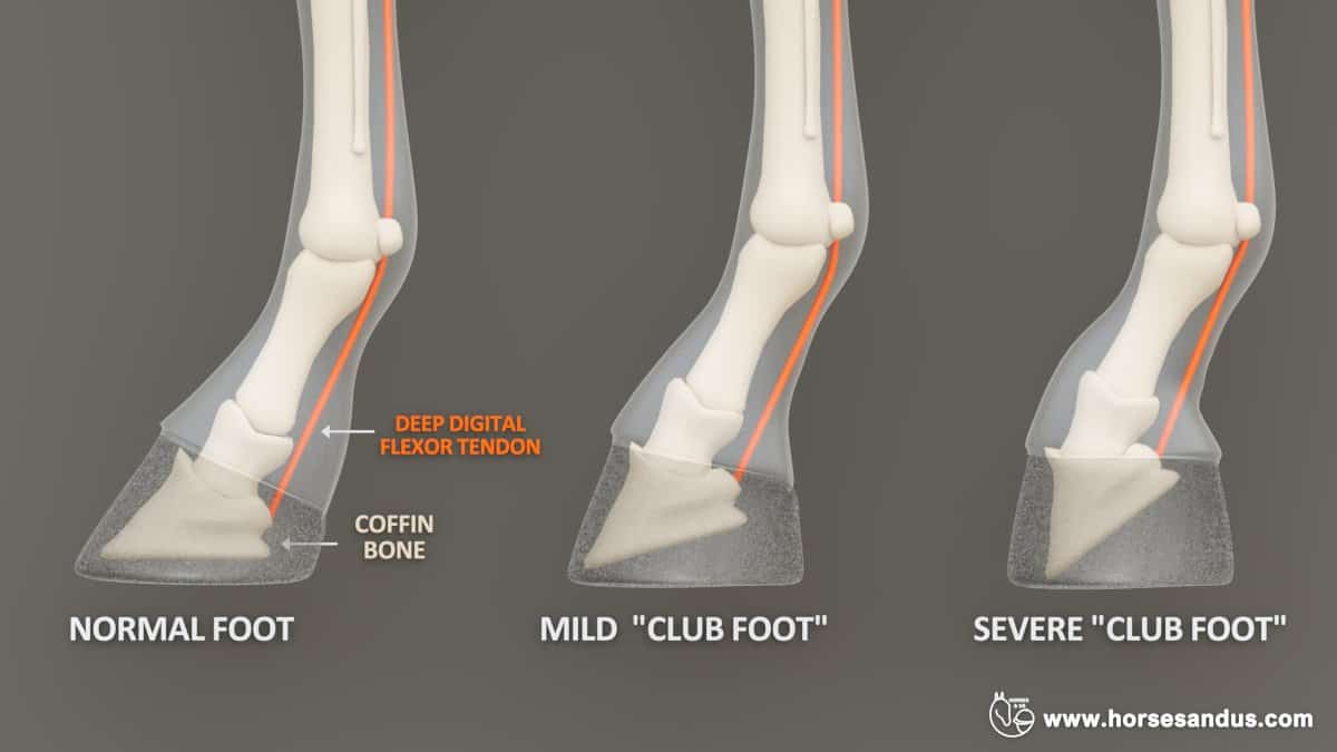
Club Foot is characterized by a very upright hoof. It is associated with the deep digital flexor tendon being much shorter than the accompanying bones. Therefore it pulls on the coffin bone and rotates it downward.
In mild cases, the hoof may appear more upright than normal. In more severe cases, the hoof has a boxy appearance, with an elongated heel and toe that curves inward.
The deformity is caused by fast bone growth without a corresponding elongation of the tendon. This results in tendons that are functionally shorter than the bones.
This condition can be congenital or acquired. The acquired cases usually appear between 1 to 6 months of age.
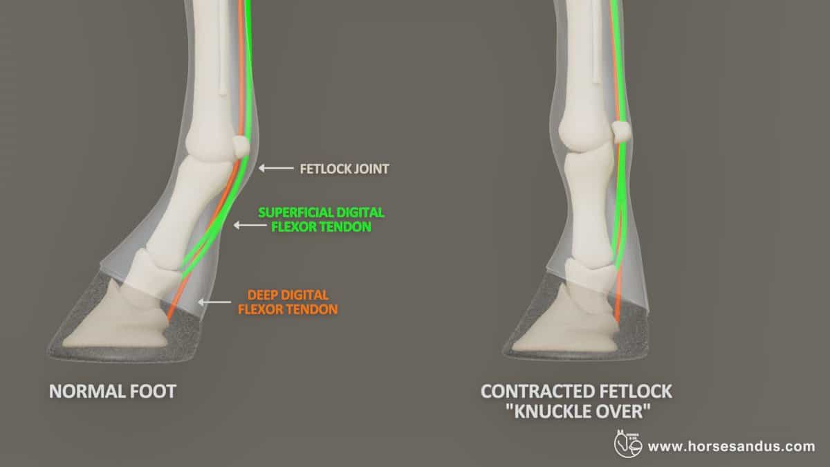
Hyperflexion of the fetlock joint is characterized by an upright fetlock and pastern, but the heel stays on the ground.
This is caused by a contraction of the superficial flexor tendon. In severe cases, the fetlock may knuckle forward.
This condition can be congenital or acquired. The acquired cases usually appear between 10 to 15 months of age.

Hyperflexion of the Carpus is characterized by the knees bending forward.
Congenital limb deformities of the carpus are quite common and can vary from very mild (where it is almost unnoticeable) to more severe (where the foal is unable to stand).
Causes of Flexural Limb Deformities in Foals
The cause of these deformities is multifactorial. The two main categories are congenital (present at birth) and developmental (acquired after birth).
Congenital– Maternal diseases or intrauterine malpositioning in an abnormally large foal relative to the mare’s size.
Developmental – Occur most commonly during fast growth times, which can be due to genetics or excessive nutrition.
Rapid bone growth sometimes may not be accompanied by a corresponding elongation of the flexor tendons, leading to the development of hyperflexion deformities.
It is also believed that pain due to rapid growth can cause contraction of the muscles and tendons.
Treatment of Flexural Limb Deformities
The treatment depends on the degree of the deformity.
Mild flexural deformities generally do not require any treatment. They can be managed by controlled exercise.
In moderate cases, the treatment consists of trying to relax the muscles and tendons. This is done with one or more of the following methods: medication, hoof trimming, shoeing, splinting, and casting.
Severe cases require surgery which consists of cutting ligaments or tendons to allow stretching.
(sources of equine flexural limb deformities used for this article: osu, umn, ker, sciencedirect )
Equine Wobbler Syndrome
Equine Wobbler Syndrome is a malformation of the cervical vertebrae (neck) characterized by the narrowing or instability of the spinal canal. This malformation causes the compression of the spinal cord, which leads to neurological symptoms in the limbs.
Horses with this disease present a wobbling or uncoordinated gait. They have a loss of sensation in their limbs and so are not aware of where the limbs are.
Equine Wobbler Syndrome is considered a Developmental orthopedic Disease in horses because it typically occurs between six months and three years due to vertebral malformations while the horse is growing.
However mature horses can also be affected due to other causes such as arthritis or injuries.
The signs of wobblers syndrome can develop gradually or have a sudden onset. The first sign is usually the lack of coordination of the hind legs. Horses can use their vision to help their front legs’ coordination, but they cannot do this with their hind legs.
Two Distinct Types of Wobbler Syndrome
Wobbler syndrome can have a dynamic or a static form. The dynamic form presents symptoms only when the neck is in a certain position. The static form presents signs regardless of the neck position.
Cervical vertebral instability. Dynamic compression of the spinal cord by the vertebrae when the neck is flexed. This type typically occurs in young horses between 4 and 18 months of age.
The location of compression is usually between the 3rd and 5th cervical vertebrae.

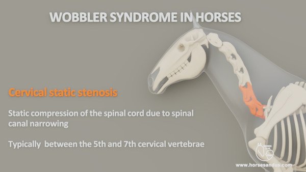
Cervical static stenosis. Static narrowing of the cervical canal. Signs manifest regardless of the neck’s position. This type typically occurs in horses between 1 and 4 years of age.
The location most commonly affected is between the 5th and 7th vertebrae.
However, the distinction between these two types is not always clear. Sometimes all vertebrae from c3 to c7 are affected by a narrowed canal.
Causes of Equine Wobbler Syndrome
The exact cause of wobbler syndrome is not clear but it can be associated to
- Genetic predisposition
- Unbalanced nutrition
- Rapid growth
- Physical trauma
It is believed that the wobbler syndrome can be related to neck length, where horses with longer necks are more likely to develop this condition.
The incidence of wobbler syndrome is higher in males than females.
Treatment for Equine Wobbler Syndrome
The options for treatment of this disease can be:
- Reducing nutrient intake (to delay rapid bone growth) and limiting exercise. This can produce good results when the condition is diagnosed early.
- Medication to decrease the swelling of the nerves and therefore the pressure.
- Surgery which involves stabilizing the affected area to allow bones and spinal canal to remodel.
(sources of equine wobbler syndrome used for this article: College of Agriculture Food and Environment, University of Kentucky Cooperative Extension Service )